Plant Anthoceros
Systematic Position
Division: Bryophyta
Class : Anthoceropsida
Order : Anthocerotales
Family : Anthocerotaceae
Genus : Anthoceros
Occurrence
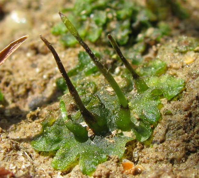
Anthoceros grows on moist soil. They are present in shady places or in the crevices of rocks. Anthoceros erecrus, A. himalayensis, and A. chambensis are commonly found in Western Himalayas.
General structure
Anthoceros has thallus body. It is lobed and it has irregular or dichotomous branches. The lobes have a wavy margin. Anthoceros form small rosette like plant. Unicellular rhizoids are attached to the underside of the thallus. Small mucilaginous cavities are present on the ventral side. These cavities contain colonies of a blue green alga like Nostoc. Stomata like small slits are present on the dorsal side of the thallus. Mucilage oozes out through these slits.
Internal structure of Anthoceros
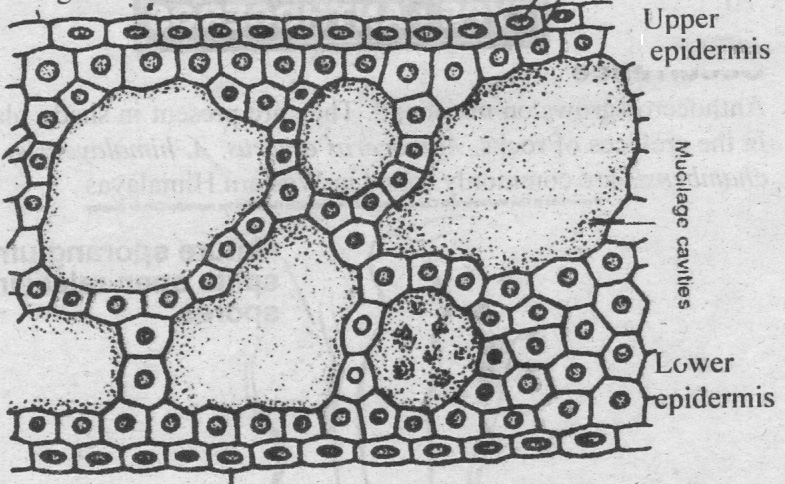
Internal Structure of the thallus
The thallus has uniform tissue of parenchymatous cells.
Epidermis is present on both sides. The cells in the upper region contain the chloroplasts. Generally each cell contains a single chloroplast. Each chloroplast has a pyrenoid. The thallus is thickest in the middle. It gradually becomes thinner towards the margins. Cells of the lower epidermis give rise to smooth unicellular rhizoids.
Growth of thallus
The growth occurs by single apical cell in initial stages. But mature plants have several scattered apical cells on the margin. So growth takes place at many points. It forms rosette like thallus.
Reproduction
Vegetative Reproduction
1. Death of older parts: Vegetative reproduction takes place by the death of older parts. Younger parts form new thallus.
2. Tuber: Some thallus forms tubers. These tubers are rich in stored fats and proteins. These tubers germinate to on the margin of the lobes. They can survive long periods of drought. Tuber detach and from new plants.
3. Gemmae: Gemmae are also produced on short stalks on the upper surface of the thallus. These are also act as vegetative reproductive bodies.
Sexual Reproduction
Anthoceros has both monoecious and dioecious species. Male plants are smaller than the female in the dioecious species. In monoecious species the antheridia are produced earlier than archegonia. The sex organs are deeply embedded in the thallus.
Antheridia
The antheridia are present on the upper side of the thallus in small cavities. They are found in groups of 2-4. The antheridial cavities are completely covered by a double layer of cells. They have no opening to the outside. Each antheridium is borne on a multicellular stalk. The main body of the antheridium is globose. It has a single celled thick jacket. Antheridia have mass of androgonial cells. They give rise to biflagellate antherozoids.
Development of Antheridium
1. Antheridium develops from a single superficial cell of the thallus. This cell divides by a transverse division into an outer and an inner cell. The inner cell acts as antheridial initial. A space is produced between inner and outer cells. This space is filled by mucilage. The inner cell is pushed towards the base of cavity. The outer cell divides to form roof of antheridial cavity.
2. The antheridial initial divides by one or two vertical divisions. It produces two or four cells. Each of which develops into an antheridium. Thus it gives rise to a group of antheridia in each cavity.
3. Each antheridial cell divides by transverse division. It produces a primary stalk cell at the base and a primary antheridial cell at the top. The primary stalk cell divides to from multicellular stalk.
4. The primary antheridial cell first divides by two vertical divisions at right angle to each other. It produces a group of four cells. One or more transverse divisions take place. It produces 2-3 tiers of four cells. Periclinal divisions take place in all these cells. It produces central androgonial cells and peripheral jacket cells.
5. The jacket cells by further divisions produce a single layered wall. The androgonial cells produce a mass of androcytes or antherozoid mother cells. The contents of each antherozoid mother cell are transformed into a single antherozoid. The roof of the antheridial cavity and antheridial wall ruptures. It releases antherozoids in the surrounding water.
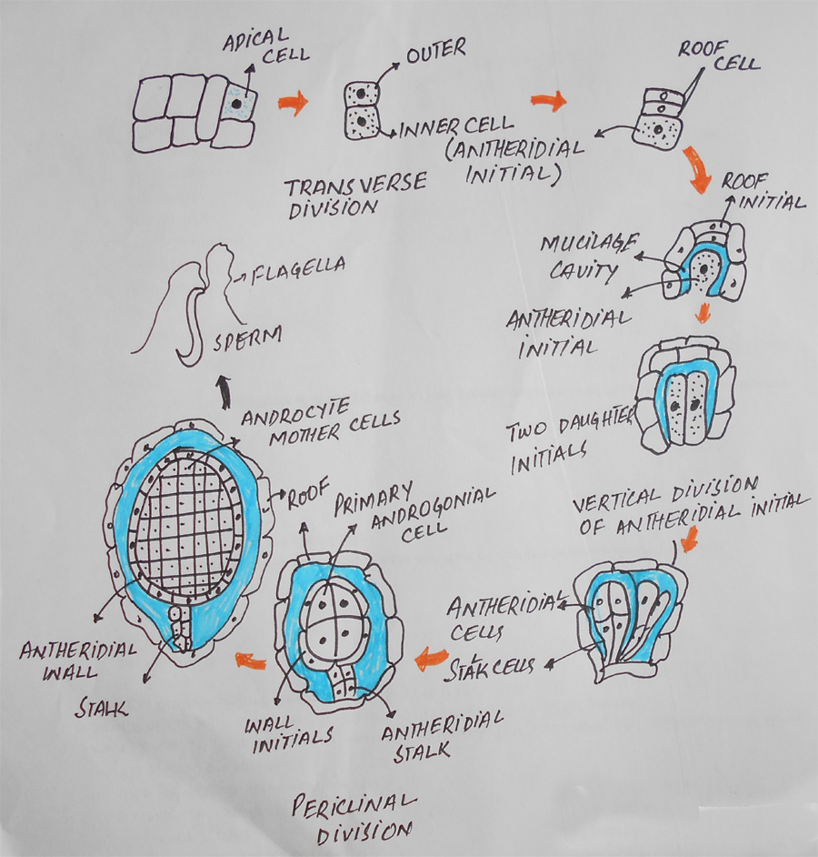
Fig: Development of antheridium
Archegonia:
Archegonia are produced close to the growing point. Archegonia are embedded in the tissue of the thallus. Each archegonium consists of an egg and a ventral canal cell four neck canal cells. The canal of the archegonium is closed at the top by four cover cells. These cells project slightly above the general surface of the thallus.
Development of Archegonium
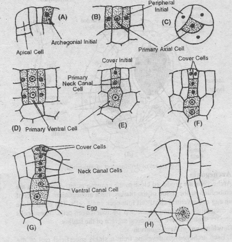
Fig: Development of Archigonium
1. Each archegonium develops from a single superficial cell of the thallus. The archegonial initial divides by three vertical divisions. It produces a large axial cell and three peripheral jacket initials.
2. The axial cell divides transversely. It cuts off a small cover cell at the top. It divides into a lower primary ventral cell and an upper primary canal cell.
3. The primary ventral cell divides transversely. It produces a larger egg or oosphere at the base and a small ventral canal cell at the top.
4. The primary canal cell divides transversely to produce t. row of four neck canal cells. The cover cell divides vertically twice to produce four cover cells. The neck canal cells and vennal canal cells produces a mass of mucilage at maturity. It forms an opening for the release of antherozoids.
Fertilization:
The plant becomes wet with dew or rain during fertilization. The antherozoids are attracted towards the archegonium chemotactically. Antherozoids enter the archegonium through the neck canal. One of them fuses with the egg to complete the fertilization. The zygote increases in size and completely fills the venter. It secretes a wall to become the oospore.
Sporophyte or Sporogonium:
The sporophyte of Anthoceros has certain unique features. Sporogonium is borne on the gametophyte. But
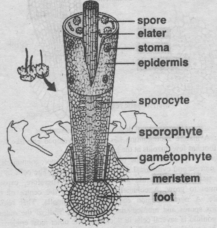
Fig: Sporophyte of Anthoceros
mature sporogonium does not totally dependent on the gametophyte.
Foot: A mature sporogonium has a well developed cup-like foot. This foot has few rhizoids at the base.
Capsule: Capsule forms the upper part of the sporogonium.
It is long, narrow and cylindrical. It has no distinct seta.
The cells in the basal part of the capsule are meristematic.
Therefore, capsule continues to grow. A columella is present in the centre of the capsule. A narrow region encircles the columella. This region contains spores and multicellular elaters.
The Wall of the mature sporogonium is several cells in thickness. The outer most epidermal layer has cutinized walls. The epidermis has small stomata with guard cells. The cells of the capsule wall contain chloroplasts. They can perform photosynthesis. Therefore, the sporophyte is not totally dependent on the gametophyte. The wall of the mature sporogonium ruptures at the apex into two valves. It exposes the columella and spores.
Development of Sporogonium
1. The oospore divides by two vertical and one transverse division. It forms eight cells. These cells are arranged into two tiers of four cells.
2. The cells of the lower tier divide to form massive foot. The foot penetrates into the tissue of the gametophyte. Certain cells on the lower surface of the foot form rhizoid like structures.
3. The upper tiers of cells divide transversely. It produces two or three tiers of cells. Periclinal divisions occur in these cells. It produces inner group of cells called endothecium and an outer group called amphithecium.
4. The cells of the endothecium divide to form columella. The cells of the columella are arranged in a square in the transverse section.
5. Cells of the amphithecium divide to produce an outer layer of jacket initials and an inner layer of sporogenous tissue or archesporium.
6. The jacket initials divides to form wall of the sporogonium. The outer most layer of jacket become epidermis. It is cutinized. Some epidermal cells divide vertically to form guard cells around a small opening. The parenchymatous cells lying below the epidermis develop chloroplasts and form the photosynthetic tissue.
7. The thickness of the sporogenous layer or archesporium is variable from single to four layered in different species. The cells of the sporogenous tissue arranged in alternate layers of larger and smaller cells. Larger cells are the spore mother cells. These cells divide meiotically to form four spores. The spores are arranged in tetrads. The smaller cells become elongated. They join together end to end and form simple or branched elaters.
8. All the cells of the sporogonium do not mature at the same time. The cells at the base of the capsule remain meristematic. They go on adding new cells towards the upper side. The new cells gradually mature into the different regions, i.e. columella, sporogenous layer and wall, of the mature capsule. In this way the sporophyte remains active for a long time. The growth at the base of the capsule continues even after its dehiscence at the top. The spores are liberated by the rupturing of the wall at the top into two valves.
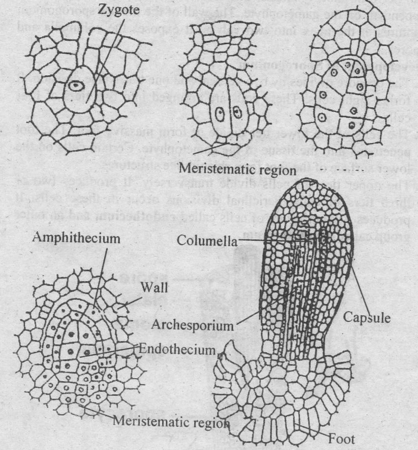
Fig: Development of Sporophyte
Spore Germination
Each spore has an outer thick wall exosporium and an inner thin wall endosporium. The spores undergo a period of rest of few weeks or months. The outer wall ruptures during germination. The inner wall protrudes out to form protonema. The cells of the protonema become green. The apical cell of protonema form thallus. Smooth rhizoids come out from certain cells of the lower surface. They fix the thallus to the soil. Some mucilaginous cavities also develop at the lower side. Nostoc filaments enter through these tubes.
Apospory
Certain cells of the sporophyte can develop into gametophytes directly under certain conditions. This phenomenon is called apospoty. It produces diploid gametophyte.
Alternation of Generation
Anthoceros shows heteromorphic alternation of generation. Thallus is a gametophyte. It develops sex organs which produce the gametes. The gametes fuse to form oospore. Oospore gives rise to the sporophyte. The sporophytes are semi-independent. The tissue of the sporophyte is diploid. The spore mother cells undergo meiosis and give rise to spores. Spores germinate to form haploid gametophyte.
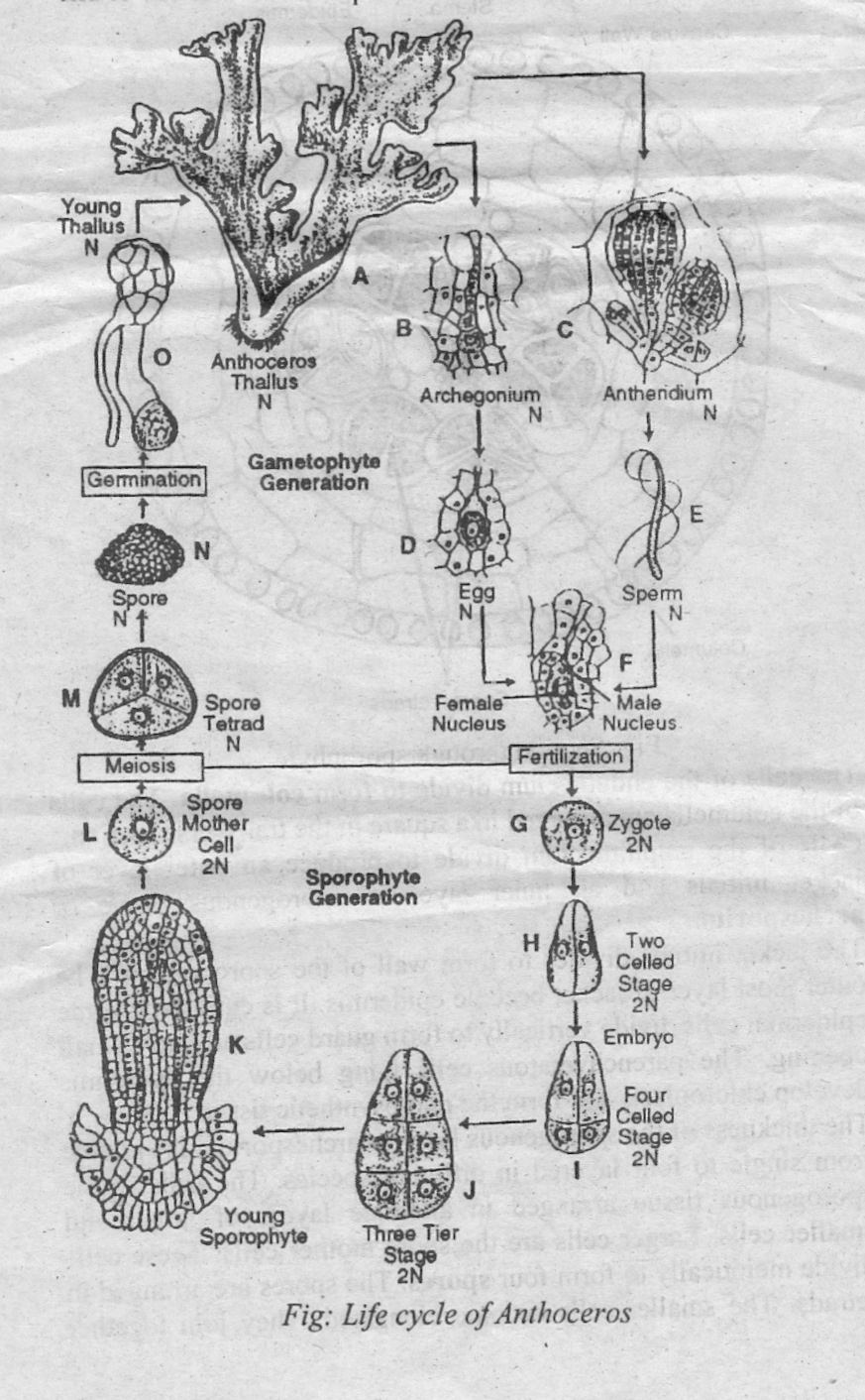
Life cycle of anthoceros