Systematic Position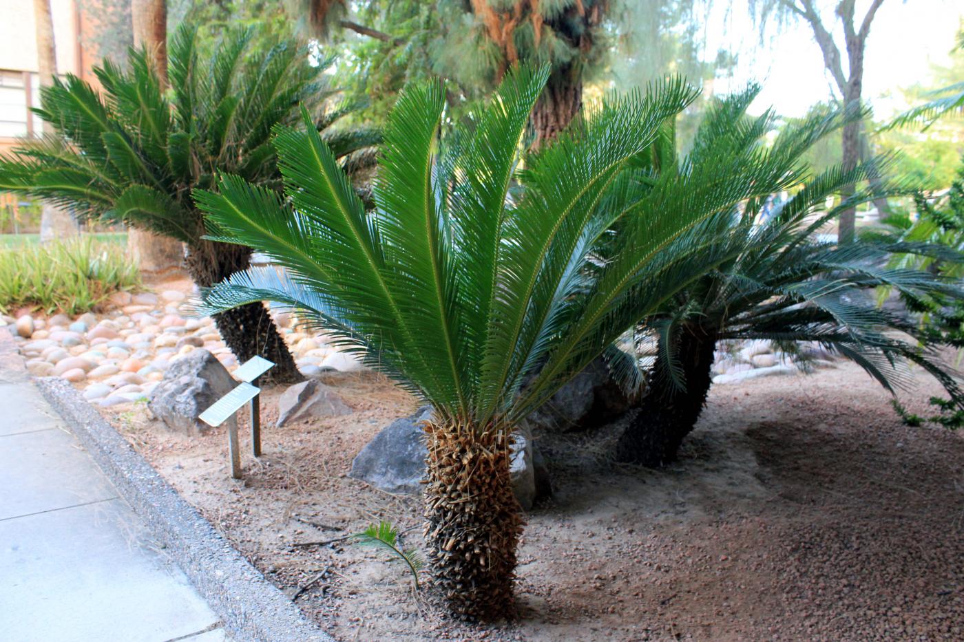
Division: Gymnospermae
Class : Cycadophyta
Order : Cycadales
Family : Cycadaceae
Genus : Cycas
Occurrence
Cycas has a limited distribution. It is found in south East Asia, South China, Southren India, South Japan and Australia. The plants grow under xerophytic conditions. Cycas is also cultivated as an ornamental plant in the gardens and parks. Cycas plants live upto hundred years or more. Cycas is called a living fossil.
Plant Body
The plant body is a sporophyte. It is differentiated into root, stem and leaves.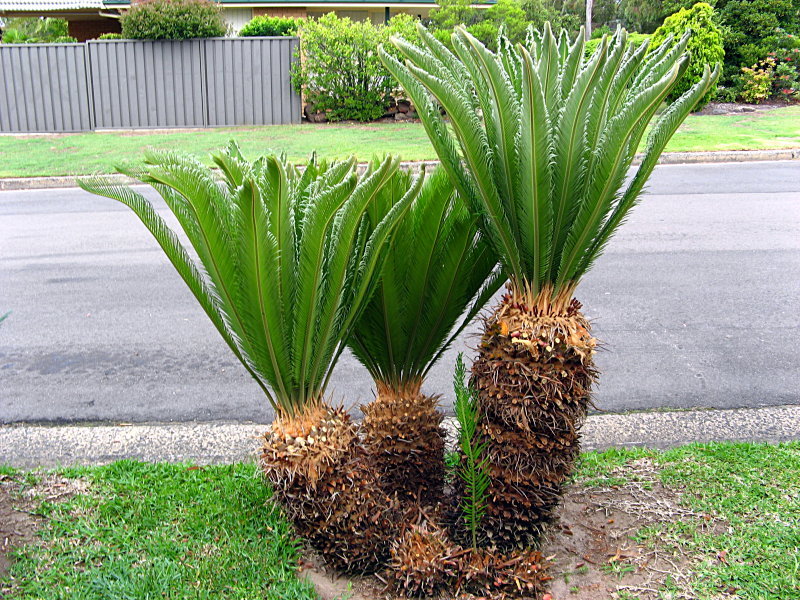
- Stem: Stem is unbranched. It is covered by thick, woody, persistent leaf bases. It makes the stem rough. The apex of the stem is ensheathed by a group of brown scales. The lower of stem is covered by pinnate compound leaves. The growth of the stem is very slow. It produces a cluster of leaves each year. Older leaves fall of after two years.
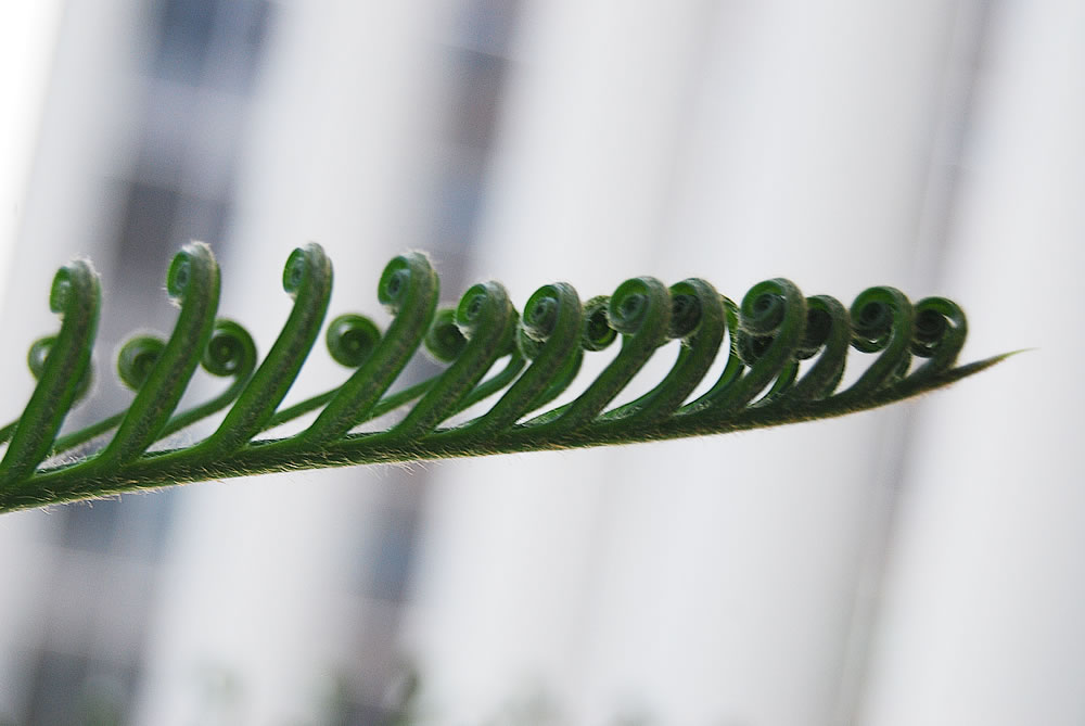
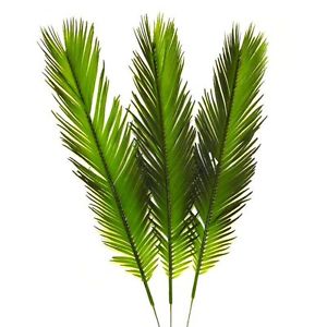 Leaves and scales: The leaves are produced in the axils of the scales near the apex. Each leaf is composed of a petiole, rachis and lateral pinnae. The young leaves show circinnate vernation. Scales are also produced each year. Therefore, the clusters of green leaves and scales alternate with each other. Scales are also persistent. Scales and leaf bases cover the surface of the old stem.
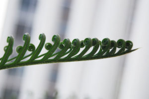
Fig: Circinate Vernation of Leaf
- Roots: The primary root persists in Cycas. It becomes tuberous. Cycas produces coralloid roots.Coralloid roots are short tufts and dichotomously branched roots. These roots contain an endophytic alga in the inner part of their cortex. Sometimes, bacteria are also present in the cortex. Bacteria fix nitrogen.
Internal Structure of Stem

Stem is internally composed of outer epidermis, cortex and vascular bundles.
- The cortex is very wide. It is composed of parenchymatous cells. Parenchymatrs tissue of the cortex contains numerous mucilaginous canals.
- Primary vascular bundles are present inside the cortex. They surround the central pith. These bundles are collateral. The primary medullary rays present between the adjacent bundles.
- Cambium is present in the form of a narrow strip in each vascular bundle. The xylem is endarch. The strength of the stem is mainly due to the presence of woody leaf bases on the surface.
- Phellogen is produced in the periphery of the cortex. It produces cork (peridium) under the persistent leaf bases.
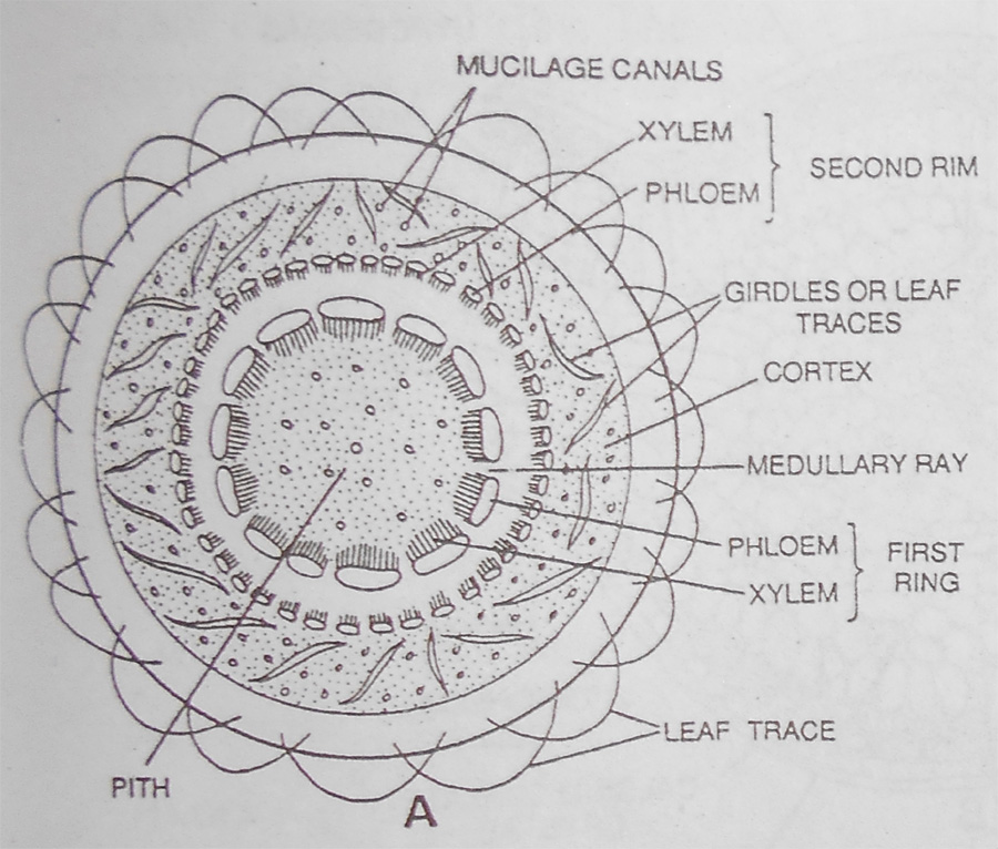
Fig: T.S of Stem of cycas
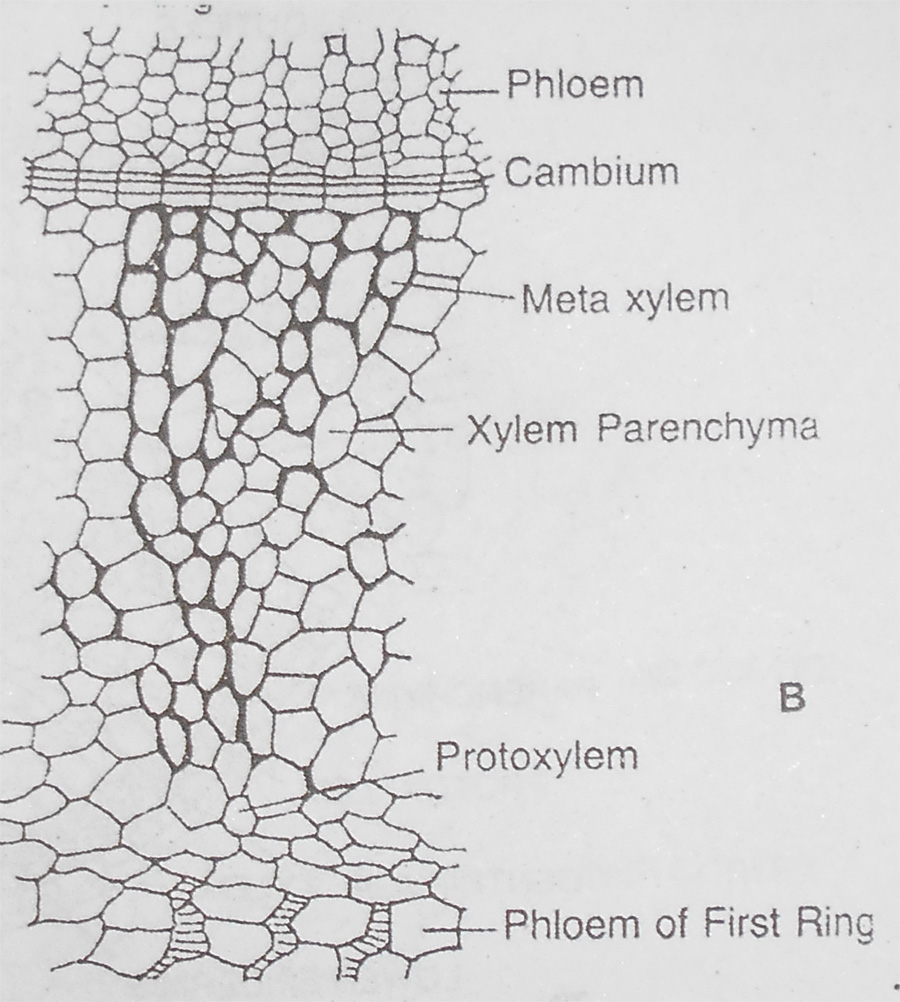
Fig: T.S of Stem
Internal Structure of the Leaf
Each pinna of leaf has typical bifacial structure in transverse section.
- Surface of leaf is covered by a single layered epidermis. The cells of the upper epidermis are slightly thick walled. But those of the lower epidermis are thin walled. Sunken stomata are present in the lower epidermis. A layer of hypodermis is present beneath the upper epidermis.
- The mesophyll is differentiated into an upper palisade layer a lower spongy parenchyma. The cells of the mesophyll are rich in chloroplasts.
- Each pinna is supplied by a single undivided vein. Transfusion tissues are present around mid ribs. They cause lateral conduction in the leaf. The vascular bundles are surrounded by pericycle and endodermis.
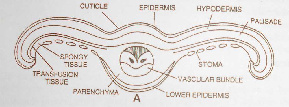
Fig A: TS of leaf of Cycas
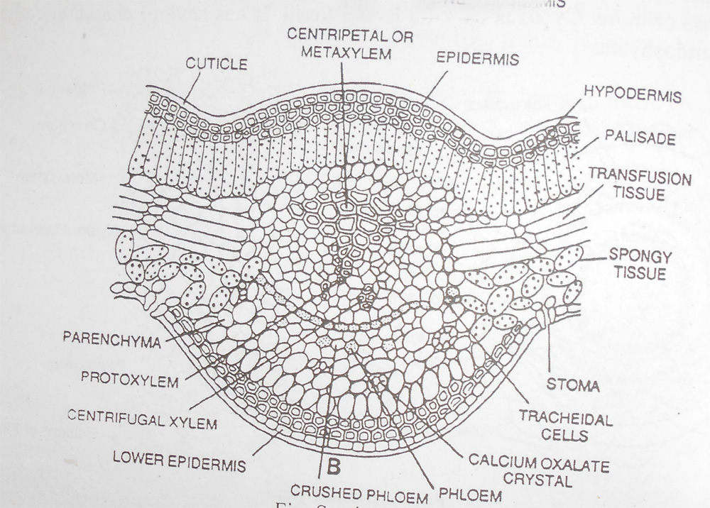
Fig B: T.S of leaf of Cycas
Internal structure of root
- Epidermis: the surface of root is covered by a single layered epidermis
- Cortex: It is many layered thick and made up of thin walled parenchymatous cells.
- Endodermis: The inner most layer of cortex is called as endodermis
- Pericycle: Thin walled cells around the vascular bundle
- Stele: The young primary root is Diarch (vascular bundles are arranged in two groups) but in older roots it become Polyarch (vascular bundles are arranged in many groups).
The xylem is exarch (Protoxylem is present towards teh outside and metaxylem towards the centre. Phloem alternates with protoxylem elements. - Pith: is present in the centre of stele. It is composed thin walled parenchymatous cells. Root cap is present at the tip of root.
Coralloid Roots
The structure is similar to that of primary root. In addition there is a conspicuous broad blue green zone in middle cortex. This is the algal zone. It lies midway between the vascular bundle and epiblemma. The cortical cells in this region get disorganized and are inhibited by blue green algae such as Nostoc and Anabaena.
In coralloid root the cortex is divided into three zones;Outer cortex, Middle cortex and Inner cortex.
The stele is diarch, triarch or tetrarch and surrounded by an endodermis which is followed by pericycle.
REPRODUCTION
Cycas is dioecious. The male and female plants are separate. Sometimes, Cycas plants also reproduce vegetatively. It produces buds on the stem. These buds grow to form new plant.
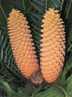
Fig: Cycas male cones
Male Cones
Male cones are produced on the male plants. Number of cones produced each year varies from one to many. Each male cone is fusiform in shape. Each cone has a central axis. It bears a number of spirally arranged microsporophyll. The microsporophylls are woody in texture. They are wedge-shaped. The microsporangia (Pollen sacs) cover the lower surface of the microsporophylls. The sporangia form sori. Each sorus has groups of two to six sporangia. Each sporophyll has several hundred sporangia. A large number of spores are produced in Cycas.
 Development of the sporangium
- Each sporangium develops from a single sporangial initial. This cell arise form the hypodermis of the sporophyll. This sporangial initial divides into an outer primary wall cell and an inner primary sporogenous cell.
- The primary wall cell divides to produce a several layered wall. The number of layers in the wall varies from four to eight. The primary sporogenous divides to produce a mass of cell called sporogenous tissue or archesporium.
- The outer most layer of the sporogenous tissue forms the tapetum. Some of the sporogenous cells increase in size and become the spore mother cells. But other cells disintegrate with the tapetum and nourish the spore mother cells.
- The spore mother cells divide meiotically to form four microspores (pollen grain).
- The sporangia become boat-shaped. Each mature microspore has two layered wall, the exine and intine. Exine is thickest at one end and becomes thinner towards the opposite end. The spores start germination before its liberation from the microsporangium.
Germination of Microspore (Pollen Grain)
The microspore cut off lateral prothalial cell towards one side of the spore. The larger cell then cuts off a small generative cell adjacent to the prothalial cell. It itself becomes tube cell. The microspore is liberated at this stage. Spores are dispersed by wind.
Female Cone
The female cones are produced on the female plants. Female cones of Cycas are very large. It is formed of megasporophylls.
Megasporophylls are loosely arranged to form crowns.
Each megasporophyll is leaf like in form.
The upper portion of the sporophyll is pinnate.
Ovules (megasporangium) are arranged in two rows in the basal half of the sporophyll.
The whole sporophyll and young ovules are covered by a dense mat of hairs.
The ovules loose this hairy covering on maturity.
Each ovule is covered by a single massive integument. It has a narrow micropyle at the tip. Integument projects around the micropyle to form a small beak. Nucellus projects into the micropyle. But later the nucellar cells in this region disorganize to form a small cavity called pollen chamber. It plays an important part in early stages of fertilization. One of the nucellar cells increases in size and becomes megaspore mother cell. It undergoes meiosis to form four megaspores. Three megaspores degenerate. Only one becomes functional megaspore.
Female Gametophyte
The megaspore (embryo sac) enlarges in size. Its nucleus undergoes many nuclear divisions. Thus several nuclei are formed in the megaspore. These nuclei occupy the peripheral region of the cytoplasm in the megaspore. Then the nuclei are surrounded by cell wall. Thus the megaspore becomes multicellular. It gives rise to the female gametophyte. The cells of the gametophyte develop numerous starch grains. The original megaspore wall persists around the prothalial tissue.
- Three cells enlarge and function as archegonial initials at the micropylar end of the prothallus. Each archegonial initial divides into tipper neck cell and a lower central cell.
- Neck cell divides to produce neck of the archegonium. The central cell enlarges. It forms the oosphere cell. The whole interior of oosphere cell is filled with dense granular protoplasm. The wall of the oosphere cell is thick. The cells of the prothallus which surround the oosphere cell form the jacket layer.
- The nucleus of the oosphere cell divides in two. The small ventral canal nucleus disintegrates. The other large nucleus becomes oosphere nucleus. It increases very much in size. The archegonium is now ready for fertilization.
- The archegonial necks open in archegonial chamber. Original megaspore wall ruptures above the archegonial chamber. The nacellar tissue below the pollen chamber disintegrates. A passage is formed between the archegonial chamber and the pollen chamber.
- A drop of mucilaginous fluid oozes out of the micropyle. The pollen grains are lodged on the micropyle. Pollen grain is trapped this mucilage and the pollen grain moves into the pollen chamber.
Male Gametophyte
The pollen grain resumes its development after pollination.
- Generative cell divides into a stalk cell and a body cell.
- Both these cells represent an antheridium.
- A pollen tube grows out of the pollen grain.
- It penetrates the nucellus. The pollen grain becomes dormant at this stage. This period lasts for about four months.
- After that the pollen grain resumes its activity.
- Two blepharoplasts appear on the two sides of the nucleus.
- These blepharoplasts develop cilia. The body cell then divides into two antherozoids.
- The antherozoid develops thousands of cilia.
- The nucleus of each antherozoid enlarged and completely fills the whole of the antherozoid. Antherozoids remain motile for several hours.
Pollination
- Pollen grain reaches the archegonial chamber by pollination.
- The wall of the pollen grain protrudes towards the archegonial chamber.
- The pollen grain bursts and release antherozoids into the archegonial chamber.
- Antherozoid enters the oosphere.
- Male nucleus unites with the oosphere nucleus.
- Fertilized oosphere secretes a thick wall and becomes the oospore.
- Oospore develops embryo.
Development of Embryo and Seed
Oospore divides into 200-300 cells. The cells of the central region disorganize to produce a cavity. This cavity is surrounded by two to three layers of cells. The cells in the lower region elongate very much to form suspensor. The cells at the tip of the suspensor develop into the embryo. The elongating suspensor pushes the developing embryo deep into the prothalial tissue. These tissues provide nutrition to the developing embryo. Whole of the nucellus is consumed during the development of the embryo. Some prothalial tissue persists in the seed and form endosperm. A thick pad of tissue develops near the micropylar end. It functions as coleorhiza. Coleorhiza protects the radical of the embryo. Ovule is transformed into the seed. Embryo has two cotyledons. They occupy the whole seed. The seeds germinate immediately. Cotyledons remain within the seed on germination. It .absorbs nutrients for the developing embryo