Occurrence
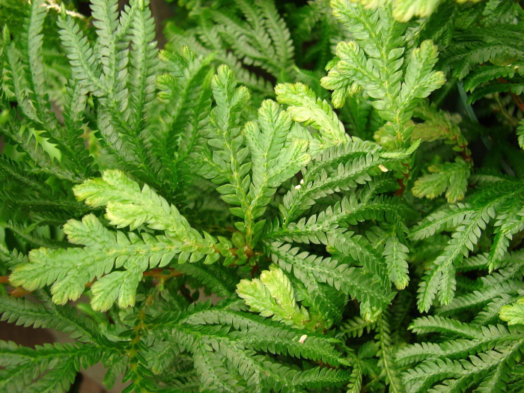
Selaginella is a tropical plant. It has world wide distribution. It grows in damp forests. Some species occur in temperate regions. They grow in moist shady places.
Selaginella_plana
General structure
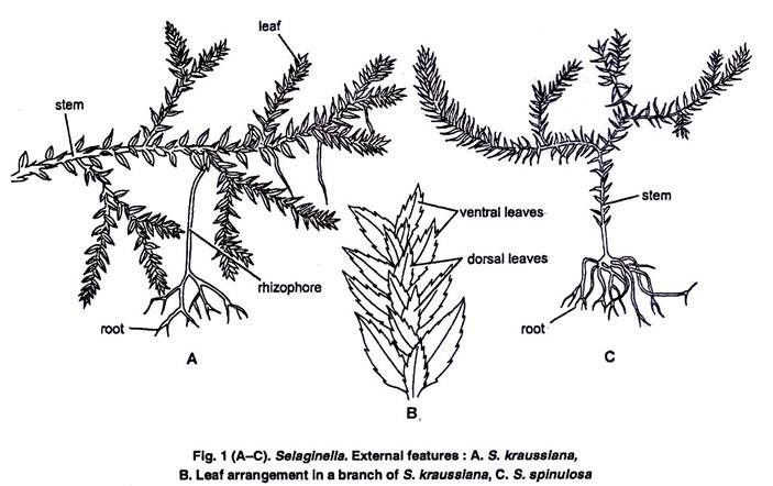
The plant body is sporophyte. The body is divided into root, stem and leaves.
Stem:
The main stem is prostrate. Some erect branches arise form the main stem.
Rhizophore:
Main stem develops leafless structures called rhizophore. Rhizophore grows downward. It develops adventitious roots at its tip. The rhizophore are intermediate in structure between the root and the stem. It is without nodes and interodes.
Selaginella
Leaves:
The main stem and the branches are covered by green leaves. Each leave has a ligule. The leaves are of two sizes, large and small. The leaves are arranged in four vertical rows. Leaves present in pairs. The larger leaf of each pair is attached toward, the ventral side of the stem and the smaller leaf towards the dorsal side. The leaves bearing sporangia in their axils are called sporophylls. Many sporophylls form cones or strobili.
Internal structure of the stem
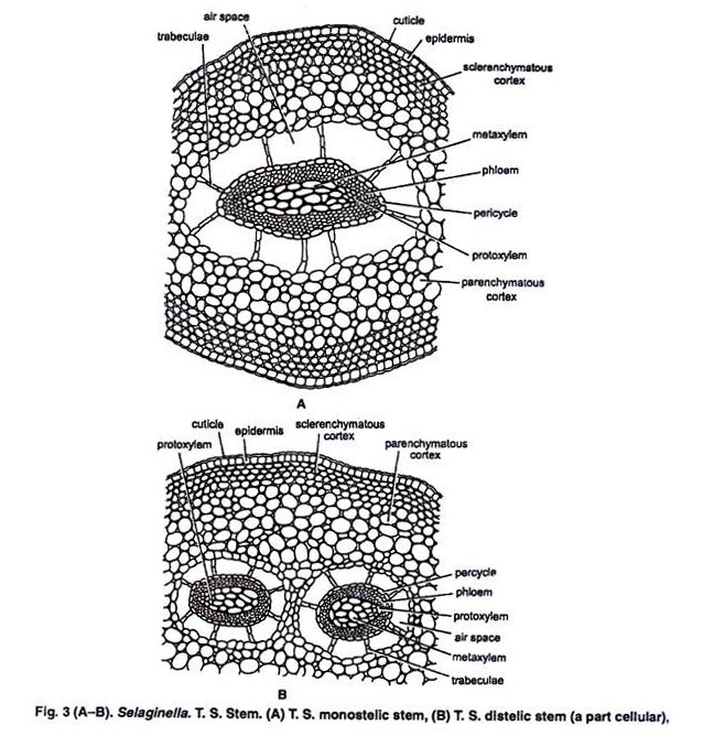
T S of Stem
In cross section, the stem is composed of epidermis, cortex and central stele.
1. Epidermis: It is outermost layer. It is without stomata.
2. Cortex: Cortex is present inner to the epidermis. It has many layered. It composed of parenchymatous cells. The cortex surrounds central stele. Cell of peripheral region of cortex contain chloroplasts. In mature regions of stem, the cortex form sclerenchymatous hypodermis.
3. Stele: Their stele is from monostelic to polystelic condition. Each stele is protostelic in nature. The metaxylem forms the solid central core. The protoxylem groups on the periphery. The xylem core is surrounded by the phloem. Outside the phloem is the pericycle. It is composed of single layer of parenchymatous cells. The stele is separated from the cortex by a wide,air space. These spaces have long radiating cells called trabeculea. Trabeculea connect the stele with the cortex.
Internal Structure of the Root:
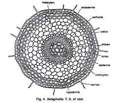
Fig: T S of Root
The root has a single layered epidermis. Inner to the epidermis is a many layered cortex. A well developed single layered endodermis separates the cortex from the stele. There is no air space surrounding the stele. The stele is protostelic and monarch. There is a single layered pericycle between the phloem and the endodermis. The internal structure of the rhizophore is similar to that of the root
Internal Structure of the Leaf:
The leaf is covered by a single layered epidermis. The cells of epidermis contain chloroplasts. Stomata are present on the upper, or on the lower, or on both sides of the leaf. The mesophyll is formed of parenchymatous cells. These cells are loosely arranged and they have numerous intercellular spaces. Each cell contains one or more chloroplasts. Each chloroplast contains several pyrenoid-like bodies. The mesophyll is traversed by a single vein.
Sporangia
Selaginella is heterosporous. The larger spores are megaspores and the smaller spores are microspores. Megaspores are produced in megasporangia and microspores are produced in microsporangia. Both sporangia are borne in the axils of leaves called microsporophyll and megasporophylls. This condition is called stachyosporous. The sporophylls form definite cones or strobili. Both kinds of sporangia are found in the same strobilus. Megasporangia are present in the basal portion and the microsporangia are present in the upper part of the cone.
Each microsporangium contains several microspores. But them are only four megaspores in each sporangium. The mature spores are pyramidal in shape. The sporangial wall consists of three layers. The inner most layer is tapetum. They provide nourishment to the developing spores. A slit is produced in mature sporangia.The spores come out of this slit.
The spores germinate to develop gametophytes. Microspore give rise to male gametophytes and the megaspores produces female gametophytes. Both male and female gametophytes remain within the walls of the spores. The young embryo develops in the megaspore. This is an approach towards the seed habit.
Internal structure of the stem
In cross section, the stem is composed of epidermis, cortex and central stele.
1. Epidermis: It is outermost layer. It is without stomata.
2. Cortex: Cortex is present inner to the epidermis. It has many layered. It composed of parenchymatous cells. The cortex surrounds central stele. Cell of peripheral region of cortex contain chloroplasts. In mature regions of stem, the cortex form sclerenchymatous hypodermis.
3. Stele: Their stele is from monostelic to polystelic condition. Each
stele is protostelic in nature. The metaxylem forms the solid central core. The protoxylem groups on the periphery. The xylem core is surrounded by the phloem. Outside the phloem is the pericycle. It is composed of single layer of parenchymatous cells. The stele is separated from the cortex by a wide,air space. These spaces have long radiating cells called trabeculea. Trabeculea connect the stele with the cortex.
Internal Structure of the Root:
The root has a single layered epidermis. Inner to the epidermis is a many layered cortex. A well developed single layered endodermis separates the cortex from the stele. There is no air space surrounding the stele. The stele is protostelic and monarch. There is a single layered pericycle between the phloem and the endodermis. The internal structure of the rhizophore is similar to that of the root
Internal Structure of the Leaf:
The leaf is covered by a single layered epidermis. The cells of epidermis contain chloroplasts. Stomata are present on the upper, or on the lower, or on both sides of the leaf. The mesophyll is formed of parenchymatous cells. These cells are loosely arranged and they have numerous intercellular spaces. Each cell contains one or more chloroplasts. Each chloroplast contains several pyrenoid-like bodies. The mesophyll is traversed by a single vein.
Sporangia
Selaginella is heterosporous. The larger spores are megaspores and the smaller spores are microspores. Megaspores are produced in megasporangia and microspores are produced in microsporangia. Both sporangia are borne in the axils of leaves called microsporophyll and megasporophylls. This condition is called stachyosporous. The sporophylls form definite cones or strobili. Both kinds of sporangia are found in the same strobilus. Megasporangia are present in the basal portion and the microsporangia are present in the upper part of the cone.
Each microsporangium contains several microspores. But them are only four megaspores in each sporangium. The mature spores are pyramidal in shape. The sporangial wall consists of three layers. The inner most layer is tapetum. They provide nourishment to the developing spores. A slit is produced in mature sporangia.The spores come out of this slit.
The spores germinate to develop gametophytes. Microspore give rise to male gametophytes and the megaspores produces female gametophytes. Both male and female gametophytes remain within the walls of the spores. The young embryo develops in the megaspore. This is an approach towards the seed habit.
Development of Sporangia
The development of micro and megasporangia is similar upto the formation of spore mother cells.
1. The sporangia initials are present in the axil of the leaf. The sporangial initials divide to form outer cells called the jacket initials, and an inner group of cells called archesporial initials.
2. The archesporial initials divides to form mass of sporogenous tissue. The outer most layer of the sporogenous tissue forms tapetum. The jacket initials by further divisions give rise to a jacket.
3. All the sporogenous cells in the microsporangia become spore mother cells. The spore mother cells separate from each other. They undergo meiosis to form microspores. Several spore mother cells are produced in the megasporangium. But only one spore mother cell is functional. All others disintegrate. The spore mother cells divide meiotically to produce four megaspores. The development of the megaspores started before their shedding from the sporangia.
Gametophytes
Development of the Male Gametophyte:
I. The development of the male gametophyte started within the microsporangia. Microspore divides into two unequal cells. The smaller cell is called prothalial cell. The larger cell is called the antheridial cell.
2. The prothalial cell does not divide further. Antheridial cell divides to produce 12 cells. Four cells occupy the centre. They become primary androgonial cells. These cells are surrounded by the remaining eight peripheral cells. The microspores are liberated from the sporangia at this 12-cell stage.
3. The outer eight cells form the jacket of the antheridium. The androgonial cells divide to produce a mass of 128-256 androcytes or antherozoid mother cells. Each androcyte develops into biflagellate antherozoid. The prothalial cell and jacket cells disintegrate and liberate the antherozoids in the surrounding water.
Development of the Female Gametophyte:
The germination of the megaspores started in the megasporangium. Spore increases in size. Nucleus of the spore undergoes several divisions. It makes the spore multinucleate. A large central vacuole develops in the spore. It pushes the whole of cytoplasm towards the pointed end of the spore. The vacuole gradually disappears. Two or three layers of cells are formed towards the pointed end of the spore. A clear membrane diaphragm separates the cellular layers from the rest of the cytoplasm. The spore wall ruptures at the pointed end exposes the cellular layers. The exposed cells develop chloroplasts.
Some cells produce rhizoids.
1. Several superficial cells of exposed tissues become archegonial initials. The archegonial initial divides into an upper primary cover cell and a lower central cell.
2. The primary cover cell divides to form the neck of the archegonium. The central cell divides to produce an upper primary canal cell and a lower primary ventral cell. The primary canal cell functions as single neck canal cell:
3. The primary ventral cell divides to produces a lower egg or oospbere and an upper ventral canal cell. The surrounding vegetative tissue forms the wall of the venter. The ventral canal cell and the neck canal cells of mature archegonia disintegrate. They form a passage for the entry of antherozoids.
Fertilization
Fertilization always takes place in moisture. Antherozoids swim in water. One antherozoid enters into archegonium. It fuses with oosphere to produce oospore.
Development of the Embryo:
1. The oospore divides into two cells. The upper cell enlarges. It is cilled suspensor. The lower cell is called the embryonal cell. It develops into the embryo. The suspensor pushes the developing embryo into the tissue of the gametophyte.
2. The embryonal cell divides to form eight cells or octants. Two cells of the octants divide more rapidly. They produce an outgrowth called foot on one side. Foot is the chief food absorbing organ of the developing embryo.
3. The remaining cells of octant form a mass of cells. The cel tral group of cells in this miss forms the apical meristem. The remaining cells of these mass produce rudiments of the first leuves or cotyledonary leaves.
4. Root primordium arises as a protuberance between the foot and the suspensor. The root primordium forms rhizcrphore.
5. Further growth of the apical meristem pushes the embryo out of the gametophytic tissue. Stem grows upward taking with it the cotyledonary leaves. The rhizophore grows downward and produces adventitious roots.
Alternation of Generation
Selaginella shows a regular alternation of sporophytic and gametophytic generations. The vegetative plant is diploid sporophyte. It produces haploid micro and mega spores by meiosis. These spores give rise to male and female gametophytes. Gametophytes produce male and female gametes. The gametes fuse to form diploid oospore. This oospore develops into the sporophyte.
Evolutionary advancement of Selaginella:
Approach to seed habit:
Selaginella shows an evolutionary advancement over the other Pteridophyta. It has an approach towards seed habit due to following advanced characteristics.
1. The production of gametes, fertilization and the development of the embryo, take place on the sporophyte. Megaspore is never released from the sporophyte.
2. Selaginella is heterosporous. The microspore produces the male gametophyte: It completes its development within the wall of the spore.
3. Megaspore contains a large amount of reserve food material. The female gametophyte completes its whole development within the megaspore wall. Fertilization and the development of the embryo also take place within spore wall. The developing gametophyte arid the embryo use the reserve food.
4. In many cases the megaspore is not released from the megasporangium. The development of the gametophyte, fertilization of the oosphere and the early development of the embryo take place while the spore is still in the sporangium.