Fig: porella-arboris-vitae
Systematic Position
Division: Bryophyta
Class : Hepaticopsida
Order : Jungermanniales
Family : Porallaceae
Genus : Porella
Occurrence:
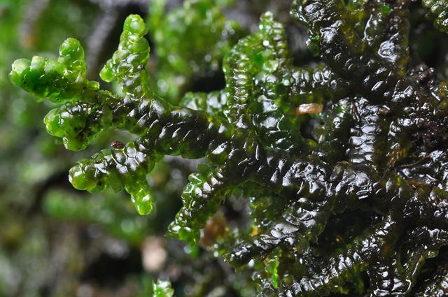
Most species of Porella are distributed in tropical to temperate regions. It grows on moist rocks, on the shaded sides of tree trunks.
It forms dense mats closely covering the ground. Some are aquatic and grow in cool waters. They are cosmopolitan.
General Structure:

Fig: A: Male plant. B: Female plant C: leaves
Plant body is gametophyte and composed of three parts
- Stem: It is slender, dorsiventral, extensively branched. Prostrate, bi or tripinnatly branched.
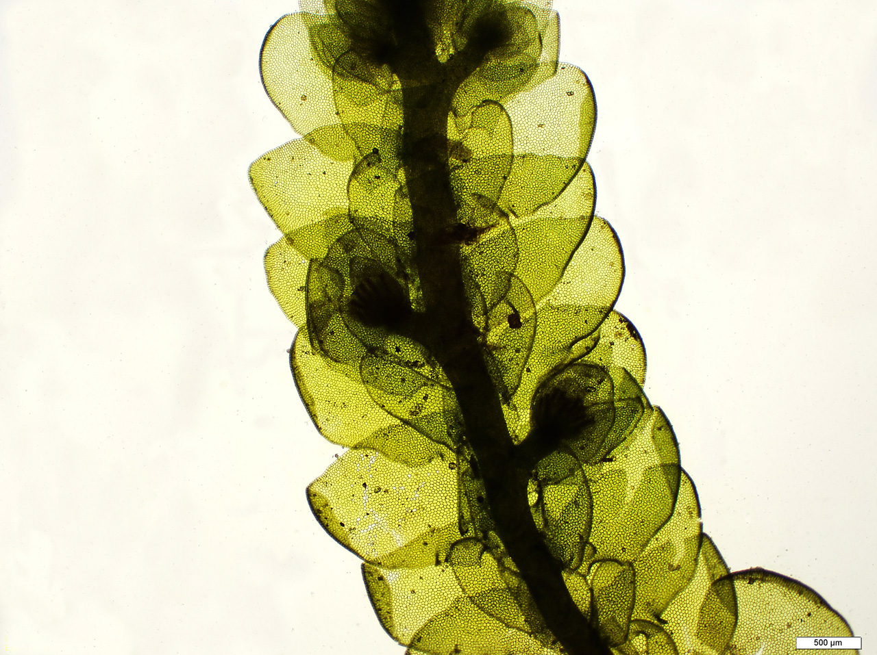
- Leaves: Three rows of leaves are present. Two of them are dorsal and a single row of small ventral leaves. The dorsal leaves are closely overlap each other and are bilobed. The two lobes are unequal. The lower lobe is smaller and called Lobule. The smaller ventral leaves are called amphigastia.
- Rhizoids: Many smooth walled rhizoids arise from the base of ventral leaves. The absorption of water takes place directly through leaves and stem cells.

Leaves of porella
Internal structure of thallus
There are similar internal tissues in stem, branches and leaves. The leavas of PoreIla are one cell in thickness. They have no mid-rib. Each cell is polygonal, uninucleate and contains several chloroplasts.
Stem shows little more internal differentiation of tissues. The axis shows two parts, i.e., outer cortex and the central medulla. The outer cortical cells are small and thick-walled. The inner medullary cells are large and thin-walled. The superficial cells of the stem give rise to paraphylls. Paraphylls are filamentous outgrowths. They supplement the photosynthetic activities of leaves.Reproduction:
a) Vegetative reproduction:
Vegetative reproduction takes place by following methods:
1. Death and decay: The basal older parts of the thallus die and decay. It separates the branches. These branches grow and become independent plants.
2. Discoid gemmae: In some species (P. rotundifolia) discoid multicellular gemmae ace produced on the lower surface of the leaves. Gemmae germinate to produce new plants
3. Tubers: These are modified special underground branches. These are formed at the end of growing season. They remain dormant throughout the unfavourable conditions.
Sexual reproduction:
The plants of Porella are mostly dioecious. Only one specie is monoecious.
(a) Male plants
The male plants are smaller. Antheridia develop on special lateral branches.
The leaves adjacent to the sex organs are called bracts. A long-stalked antheridium is present in the axil of each bract. Each antheridium consists of a globular body or capsule and a long slender stalk. A large number of spermatozoids are present within the wall of capsule.
Development of antheridium
1. The antheridial initial lies close to the growing apex of the male branch, This cell enlarges. It undergoes transverse division to form basal cell and upper cell.
2. The basal cell forms embedded portion of stalk. The outer cell acts as antheridial mother cell. It divides transversely into outer primary antheridial cell and lower primary stalk cell.
3. The primary stalk cell forms stalk. The primary antheridial cell divides vertically. It forms two cells. These cells divide to form four jacket initials and two primary androgonial cells. The jacket initials divide to form wall of antheridium.
4. The two primary androgonial cells divide to form a large mass of androgonial cells. These cells divide to form androcytes mother cells.
5. Each androcyte mother cell finally produces two androcytes. Each androcyte is changes into coiled biflagellate sperm.
6. The jacket bursts due to swelling of the mass of androcytes. The antherozoids are then liberated from the androcytes (sperm cells) in water. The sperms swim in water and reach the archegonia.
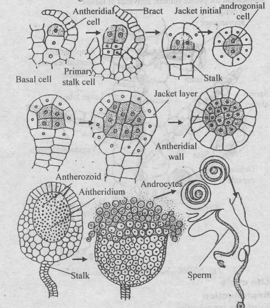
Fig: Stages of development of antheridia
Female plants
Female plants are larger than the male. Archegonia arc always present at the apex of an archegonial branch. Archegonia are present in clusters. The female branches have fewer bracts. The archegonia and bracts forms structure called involucre. Campanulate perianth is present inside the involucre. This perianth is divided into a number of lobes. The rosette, involucre and perianth form a cup shaped structure at the top of female shoot.
The archegonium consists of neck and the venter. The neck consists of five vertical rows of neck cells. These cells enclose 6-8 neck canal cells.
Development of archegonium
1. The archegonium develops from the apical cell. Each archegonial initial enlarges. It divides into basal cell and outer cell.
2. The basal cell divides to form the stalk of the archegonium. Outer cell acts as archegonium mother cell. It divides to form two large peripheral cells and one small primary axial cell.
3. The two large peripheral cells divide to form a ring of five peripheral cells. The five peripheral cells divide to form neck and venter. The primary axial cell divides transversely. It forms upper primary cover cell and lower central cell.
4. The primary cover cell forms four cover cells. The central cell divides to form upper primary neck canal cell and lower primary venter cell.
5. Primary neck canal cell divides to form 6–8 neck canal cells.
Primary venter cell divides to form upper small venter canal cell and lower large egg cell.
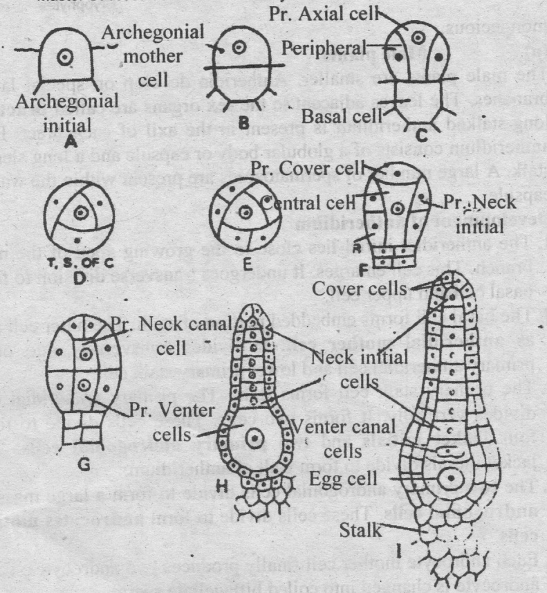
Fig: Archigonial development
Fertilization:
The neck canal cells and the venter canal cells of mature archegonia degenerate. They form mucilage-like substance. It exerts pressure and burst the cover cells. The mucilaginous fluid oozes out of ruptured neck and attracts the sperms. A large number of sperms collect near the neck. But only one of them passes down and fuses with the egg to form the oospore.
Sporophyte
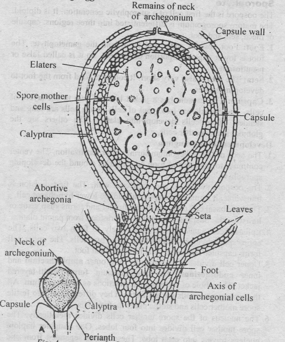
Fig: Sporangium
The oospore is the first cell of a sporophytic generation. It is diploid. The mature sporogonium is differentiated into three regions: capsule or theca, seta and foot.
1. Foot: Foot absorbs water and food from the gametophyte. The
foot is lower expanded portion of the seta. So it is called false or pseudofoot.
2. Seta: The seta is also very short. It conducts food from the foot to developing spores.
3. Capsule: The capsule has a jacket layer of two to six layers of cells. The capsule has two types of cells. Some cells are long and slender. These cells develop into elaters. The others are the globular spore mother cells.
Development of sporangium
1. The neck of the archegonium shrinks after fertilization. The venter continues to growth and form an envelope around the developing sporophyte.
2. The oospore swells up and secretes a wall. The first division is transverse. It forms upper epibasal and the lower hypobasal cell.
3. The hypobasal cell does not divide further and forms suspensor or haustorium. Suspensor is used to absorb food from parent plant.
4. Epibasal cell again divides transversely to form two cells. The entire sporophyte developed from these two cells. The upper cell forms capsule. The lower cells form seta and foot.
5. Upper cell divides many times to form outer amphithecium and inner endothecium. The amphithecium forms multi-layered jacket. The whole of endothecium functions as archesporium. All the sporogenous cells are not fertile. They differentiate into fertile spore mother cells and sterile elater mother cells.
6. The nucleus of the spore mother cells divides meiotically. The spore mother cell divides into four lobes. One daughter haploid nucleus travels into each lobe. These spores separated from one another and form a spore-tetrad.
7. The elater mother cells form elaters. The elaters are long and narrow. They are scattered in between spores.
The mature capsule is covered by calyptra. The capsule opens by four valves along four lines. The valves separate and bend back upon themselves. So spores and elaters become free.
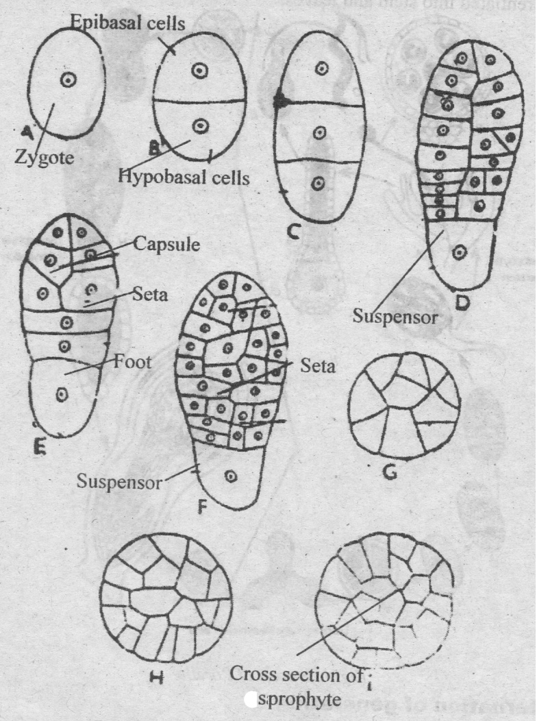
Fig: Development of Sporangium in Porella
Germination of spore
The spores are haploid. They are first cell of gameiophytic generation. The spore germinates to produces a short protonema. The protonema may be single celled or filamentous. The terminal cell of the protonema divides in all direction forming a mass of cells. One of the cells in the mass functions as an apical cell. It produces a Maltose plant body. This body produces foliose plant which is A differentiated into stem and leaves.
Alternation of generation
Porella shows heteromorphic alternation of generation.
1. Gametophyte: The plant body is gametophytes. Gametophyte is haploid. It develops antheridia and archegonia. Antheridia oroduce antherozoids and archegonium produces egg. Antherozoids fuse with egg to produce diploid oospore.
2. Sporophyte: The oospore is the first stage of sporophyte generation. It is diploid generation. Sporophyte has three parts: foot, seta and capsule. Haploid spores are produced in the capsule by meiosis. Spore is the first stage of gametophyte. Spores germinate to produce protonema stage. It gives rise to mature gametophyte completing the life cycle.
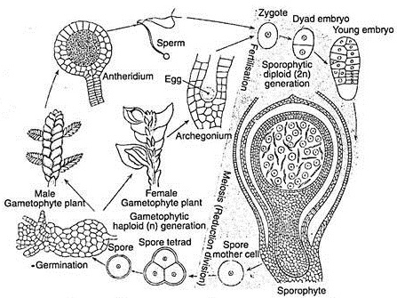
Fig: Life cycle of Porella showing alternation of generation