Conidia:
The asexual spores or conidia of ascomycetes are remarkably diverse in form, structure and modes of dispersal, but their development or conidiogenesis occurs in a limited number of ways. The cell from which a conidium develops is the conidiogenous cell and usually one or more such cells are borne on a stalk, the conidiophore. Conidiophores which are narrow and not differentiated from the vegetative mycelium are said to be micronematous (Gr. nem=a thread) whilst those that are clearly differentiated are macronematous. Conidiophores frequently arise singly as in Eurotium repens, Emericella nidulans and in many species of Penicillium. However, in certain fungi the conidiophores may aggregate to form a conidioma. Descriptive terms have been given to different types of conidioma. Conidiophores aggregated into parallel bundles (fascicles) are termed coremia (Gr. korema= a brush) or synnemata (Gr. prefix syn= together). Examples are Penicillium claviforme and Cephalotrichum (Doratomyces) stemonitis.
In many ascomycetes the conidiophores develop on or in a stroma (Gr. stroma=bed, cushion), an aggregation of pseudoparenchymatous cells. A good example of a conidial stroma is seen in the wood-rotting candle-snuff fungus, Xylaria hypoxylon.

Fig: Perithecium

Sporodochium
Here, powdery white conidia develop at the tips of the branches of the conidial stroma and, later, asci develop in flask-shaped perithecia at the base of the old stroma. The term sporodochium (Gr. spora= a seed; doche= a receptacle) is used for the cushion-like conidiomata bearing a layer of short conidiophores. An example is the conidial (Tubercularia) state of Nectria cinnabarina. Another type of conidioma is the acervulus (Lat. acervulus= a little heap), a saucer-shaped fructification which may develop inside the tissues of a host plant or may be superficial. Subepidermal acervuli develop from a pseudoparenchymatous stroma, and as the acervulus matures the overlying epidermis of the host becomes ruptured to expose conidia formed from conidiogenous cells lining the base of the saucer.
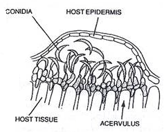
Fig: Acervulus
The conidia are held together in slime and are chiefly dispersed by rain splash. A good example of an acervular fungus is Colletotrichum. The teleomorphs of Colletotrichum, where known, are species of Glomerella, many of which are serious plant pathogens. In many ascomycetes and their allies, the conidia are borne inside flask-shaped conidiomata termed pycnidia (Gr. diminutive of pyknos=dense, packed, concentrated). Traditionally, fungi with pycnidial and acervular states have been grouped together in the artificial taxon coelomycetes , in contrast to hyphomycetes in which the conidiogenous cells are exposed on single conidiophores or in synnemata, coremia or sporodochia. Pycnidia may be superficial or embedded in host tissue. The opening of the pycnidium is generally by means of a circular ostiole. Conidia formed from conidiogenous cells lining the inner wall of the pycnidium are held together in slimy masses
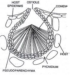
Fig: Pycnidium
which ooze out through the ostiole, sometimes as spore tendrils. They are generally dispersed by splash or in water films. In some cases the pycnidia, instead of producing conidia with an asexual function, produce spermatia which are involved in fertilization. Examples of fungi with pycnidial anamorphs are Leptosphaeria acuta with a Phoma anamorph, and Phaeosphaeria nodorum (anamorph Stagonospora nodorum;