The flagella of the green alga Chlamydomonas have been used as a model of flagellar structure. Flagella structure has been highly conserved throughout evolution, images from Chlamydomonas are virtually indistinguishable from flagella (or cilia – a term for a short flagellum) of mammalian cells including human sperm and certain epithelia. Chlamydomonas has been chosen because of the ease of growing the organism and because the flagella can be detached from the cells by pH shock or blending. Since the flagella are not essential for viability of the cell, it is relatively easy to isolate mutations affecting flagella synthesis by the cells.
A flagellum consists of an axoneme of nine doublet microtubules that surround two central microtubules, with all of the microtubules encased in the plasma membrane.
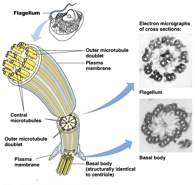
Internal structure of flagella
The flagellar membrane may have no hairs (mastigonemes) on its surface (whiplash or acronematic flagellum) or it may have hairs on its surface (tinsel or hairy or pantonematic or Flimmergeissel). There are two types of flagellar hair.
A: Non-tubular flagellar hairs made up of solid fibrils 5–10 nm wide and 1–3 m long that are composed of glycoproteins. These hairs are flexible and wrap around the flagellum increasing the surface area and efficiency of propulsion.
B: Tubular flagellar hairs about 2 m long composed of three regions:
(1) a tapering basal region 200 nm long attached to the flagellar membrane, (2) a micotubular shaft 1 m long, and
(3) a few 0.52 m-long terminal filaments
Arrangement
Algal cells can have different arrangements of flagella.
If the flagella are of equal length, they are called isokont flagella
if they are of unequal length, they are called anisokont flagella;
and if they form a ring at one end of the cell, they are called stephanokont flagella.
Heterokont refers to an organism with a hairy and a smooth flagellum.