Division : Pteridophyta
Class : Pteropsida
Order : Polypodiales
Family : Polypodiaceae
Genus : Polypodium
DISTRIBUTION:
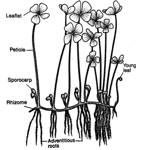
Marsilea is world-wide in distribution. Most abundantly found in tropical parts of world. The species of Marsilea are generally aquatic or amphibious in nature with their roots embedded in mud or damp soil.
GENERAL STRUCTURE:
The vegetative plant is a sporophyte. It is differentiated into roots, rhizome and leaves.
Stem:
The stem is long and slender.
Rhizome: is freely-branched underground of indefinite growth that grows on or just below the soil surface. It is differentiated into nodes and internodes.
Stolon: If stem is creeping on the surface of soil.
Leaves: The leaves are compound. Each leaf has a long petiole and four leaflets (quardifoliate). Each leaflet is triangular. Stomata are located on the dorsal side and ventral side of the leaflets.
INTERNAL STRUCTURE OF THE RHIZOME:
Internally the rhizome is composed of epidermis, cortex and central stele.
- Epidermis: The outermost single layer of cells is epidermis, composed of compactly arranged thick-walled cells and are devoid of stomata.
- Cortex:
It is differentiated into three regions
Outer cortex: it is parenchymatous with large air spaces.
Middle cortex: it is sclerenchymatous.
Inner cortex: it is solid tissue of several cell layers thick.
Endodermis:
It is present inner side of the cortex.
Stele:
Is of amphiphloic (phloem is on both sides of ring shaped xylem) siphonostele. Both the inner and outer phloem are covered by Pericycle which is single layered. It has pith in the centre. Protoxylem groups are exarch in position.
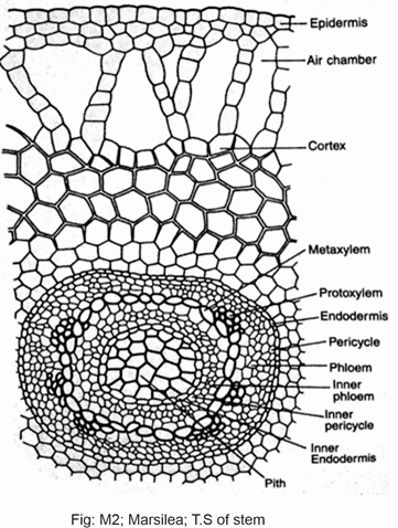
Fig; Marsilea stem internal structure
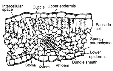
LEAF:
Both surfaces of the leaf are bound by epidermis.
Epidermis is covered by cuticle. It has sunken stomata. In hydrophytic species with floating leaves the stomata are restricted to the upper epidermis only.
Mesophyll cells are present between both epidermises. Mesophyll cells are differentiated into palisade and spongy cells.
Vascular bundle: Single vein passes through each leaf. They are concentric in nature. Each bundle has a central core of xylem surrounded by phloem. Externally vascular bundle is bound by endodermis.
REPRODUCTION:
Marsilea plant is heterosporous. The micro and megasporangia are borne in special bean shaped bodies called the sporocarp.
SPOROCARP:
The sporocarps are bisporangiate and are borne on short or long stalks called the peduncles or pedicels.
Both types of sporangia (micro and mega) are found within the same sorus.
Wall of the sporocarp: Wall is thick and three layered.
The sori: These are elongated and arranged in two alternating rows in the cavity. The wall of each sorus is formed by its own indusium. Each sorus contains a row of megasporangia and several microsporangia.
A large placenta is produced on the inner side of the wall in the young sporocarp. The placenta of two sides alternate with each other. Megasporangia and microsporangia are produced on the same placenta. Megasporangia mature earlier than the microsporangia. Each megasporangium contains a single megaspore on maturity. But each microsporangium contains several (32-64) microspores. All the tissues except indusia gelatinized in mature sporocarp.
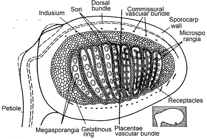
Fig: Internal structure of sporocarp showing sporangia and indusium
DEVELOPMENT OF THE SPORANGIUM:
Sporangial initials are present on the placenta or receptacle of sporocarp.
The sporangial initials present at the tip of receptacle develops into megasporangia. The initial present on the sides of the receptacle develop into microsporangia. The development of both sporangia is similar in both cases.
- The sporangial initial cuts off a jacket initial to the outside and itself becomes the archesporial initial.
- The jacket initial divides to produce single layered wall of the sporangium.
The archesporial initial cuts off two tapetal cells. These tapetal cells divide to produce two layered tapetum inner to the wall.
- The archesporial initial then divides producing 12-16 spore mother cells. Each spore mother cell undergoes meiosis and produces four spores.
- The tapetal cells provide nourishment to young spore. So they disintegrate during the development of spores.
- In the megasporangium only one spore develops further. All others disintegrate forming a mucilaginous mass or plasmodium. This mass provides nourishment to the developing megaspore. In the microsporangium all the spores develop into 32-64 microspores.

Fig: Marsilea stages of development of gametophyte
Fig; Stages of development of female gaemtophyte
DEVELOPMENT OF THE MALE GAMETOPHYTE:
The microspore germinates to produce male gametophyte. It completes its whole development within the wall of the microspore.
- The microspore divides in to two cells. The smaller cell becomes prothalial cell. The larger cell divides in two antheridial initials.
- Each antheridial initial divides to form three jacket cell and single androgonial initial. The androgonial initial of each antheridium divides to produces 16 androcytes (antherozoid mother cells). Each androcyte changes into antherozoid.
- The antherozoids have many coils and single flagella. The prothalial cell and the jacket cells of both the antheridia disintegrate.
Thus the antherozoids become free in the surrounding water.
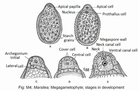
DEVELOPMENT OF THE FEMALE GAMETOPHYTE:
Each mature megaspore has a dome shaped projection or beak at one end.
The nucleus of the megaspore lies, in this beak region. It is surrounded by dense granular cytoplasm.
- Megaspore divides in to two cells. The larger cell does not divide further.
- This smaller cell functions as an apical cell. The apical cell then functions as an archegonial initial.
- 3. This archegonial initial divides to produce a small primary cover cell at the top and a central cell at the base.
- The cover cell divides to form four neck initials. They divide to form a neck. The central cell divides to produce a small upper primary canal cell and a lower larger primary ventral cell.
- The primary canal cell divides to produce two neck canal cells. The primary ventral cell divides to produce a lower larger oosphere (egg) and an upper smaller ventral canal cell.
- The ventral canal cell and the neck canal cells of mature archegonium disintegrate. It forms an opening for the entry of antherozoids.
FERTILIZATION:
Each megaspore is enveloped by a layer of mucilage. One of these antherozoids enters the archigonium fertilize the egg to produce oosphere.
Development of the Sporophyte
The oospore divides to produces four cells. Two sister cells develop stem and cotyledons.
The other two cells develop into foot and root. The vegetative cells of the gametophyte form a calyptra. The surface cells of the calyptra produce long rhizoids. The root enters the soil. Cotyledon expands to form the first simple leaf. Primary root is replaced by adventitious roots. The stem grows horizontally on the soil and form the rhizome.
ALTERNATION OF GENERATION:
Vegetative plant of Marsilea is a diploid sporophyte. It is heterosporous. It produces mega and microspores by meiosis. The spore germinates to form haploid gametophyte. The gametophyte of Marsilea is dioecious. The microspores give rise to the male gametophyte. The megaspore gives rise to the female gametophyte. Both male and female gametophytes complete their development within the spore walls. Both gametophytes produce male and female gametes. Gametes fuse to form diploid oosphere. The oospore develops into the sporophyte again.
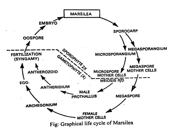
Fig; marsilea flowsheet diagram