Mitosis is a type of cell division in which one cell (the mother) divides to produce two new cells (the daughters) that are genetically identical.
The mitosis is a part of somatic cell division which includes the division of the nucleus (called mitosis or karyokinesis) and the division of the cytoplasm (called cytokinesis). The type of cell division, which ensures the same number of chromosomes in the daughter cells as that in the parent cells.
Phases of Mitosis:
Mitosis is a continuous process, but for convenience of study it may be divided into two phases:
1.Karyokinesis: This involves the division of nucleus.
2.Cytokinesis: This involves the division of cytoplasm to complete the process.
1.Karyokinesis:
Karyokinesis is the division of nucleus.
Division of Centrioles:
At the beginning of the process, the partition of the centrioles take place. That have been duplicated during interphase but were in the same centrosome. Early in the mitosis the two pair of centrioles separate and migrate to opposite sides of the nucleus, establishing the bipolarity of the dividing cells.
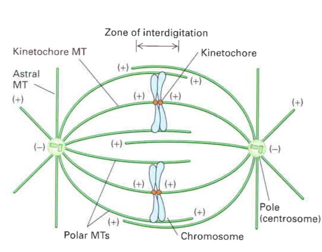
Fig: Mitotic Apparatus
Mitotic Apparatus
Three sets of microtubules originate from each pair of centrioles:
- Astral microtubules: These radiate outward and form asters.
- Kinetochore microtubule: These fibers attach to chromosomes at kinetochores.
- Polar microtubules do not interact the chromosomes, but they interdigitate with polar microtubules from the opposite pole.
These microtubules are composed of tubulin protein and traces of RNA. This specialiazed microtubule structure including asters, centrioles and spindle fibre is called mitotic apparatus.
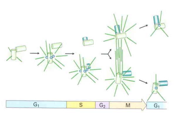
Fig Relation of centrosome duplication to the cell cycle. After the pair of parent centrioles (green) separates slightly, a daughter centriole (blue) buds from each and elongates By Gr, growth of the daughter centrioles is complete, but the two pairs remain within a single centrosomal complex Early in mitosis, the centrosome splits, and each centriole pair migrates to opposite sides of the nucleus The amount of pericentriolar material and the activity to nucleate microtubule assembly increases greatly in mitosis In mitosis, these MTOCs are called spindle ooles
STAGES OF KARYOKINESIS
Karyokinesis can further be divided into Prophase, Metaphase, Anaphase and Telophase. During interphase of the cell cycle, the chromosomes are not visible even with electron microscope, but using histological strains for DNA, a network of very fine threads can be visualized. This network is called chromatin.
- PROPHASE: (Greek; Pro- before)
(i) It is the first and the longest phase in the mitotic cell division.
(ii) Chromosomes become visible in the nucleus as short, thick, helically coiled threads.
(iii) Each chromosome has two chromatids that are joined at the centromere.
- Nuclear membrane starts dissolving.
- Nucleolus also starts dissolving and disappearing.
- Chromosomes become shorter and thicker.
Chromosomes become more and more thick ultimately each chromosome is visible having two sister chromatids, attached at centromere. Towards the end of prophase nuclear envelope disappear and nuclear material is released in the cytoplasm, nucleoli also disappear. Mitotic apparatus is organized. Cytoplasm becomes more viscous.
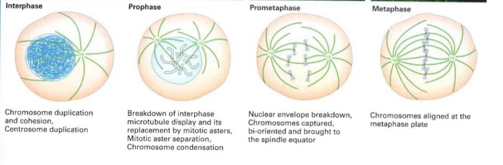
- METAPHASE (Greek Meta- between)
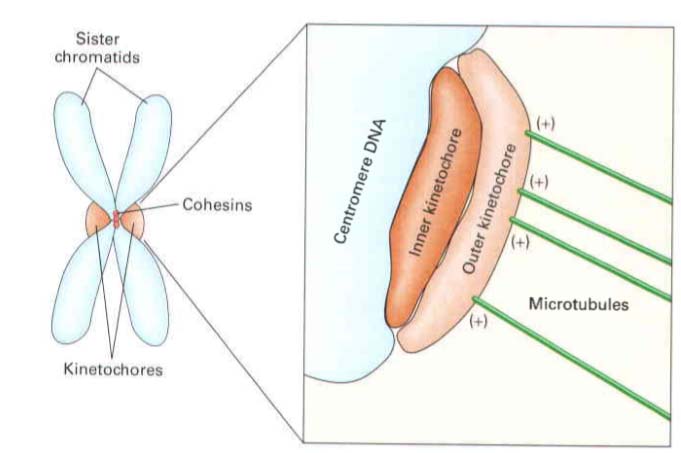
(i) Each metaphase chromosome is a duplicated structure, which consists of two sister chromatids, attached at a point called centromere or primary constriction.
(ii) The centeromere has special area the kinetochore, with specific base arrangement and special proteins where kinetochore microtubules of mitotic apparatus attach.
(iii) The kinetochore microtubules of spindle attach to the kinetochore region of chromosomes, and align them at the equator of spindle forming equatorial plate or metaphase plate.
(iv) Each kinetochore gets two fibers one from each pole.
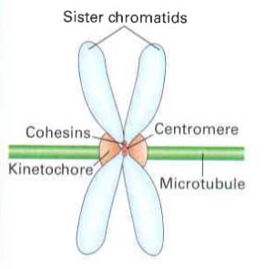
Fig: Part of condensed chromosome in mitosis. the duplicated chromosome has two sister chromatids (each with the single replicated DNA duplex), held together by cohesins at constricted region called centromere. The centromere is also the site where kinetochore forms which makes attachment to kinetochore microtubles.
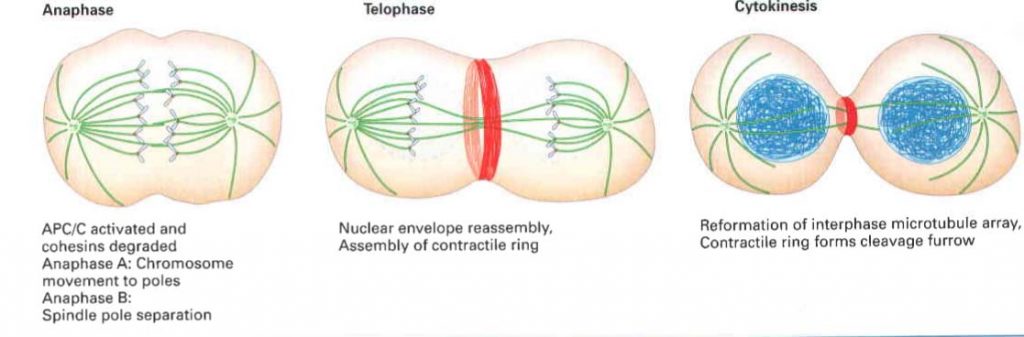
- ANAPHASE: (Greek; Ana- back)
(i) It is the most critical phase of mitosis, which ensures equal distribution of chromatids in
the daughter cells.
(ii) The kinetochore microtubules of spindle contract towards their respective poles, at the same time polar microtubule elongates, exert force and sister chromatids are separated from centeromere.
(iii) As a result half sister chromatids travel towards each pole.
4.TELOPHASE:
(i) The chromosomes reach at opposite poles.
(ii) The chromosomes de-condense due to unfolding, ultimately disappear as chromatin.
(iii) Mitotic apparatus disorganizes, nuclear membrane and nucleoli reorganize, forming two nuclei at two poles of the cell.
Cytokinesis in Plants
During mitosis two daughter nuclei are formed by karyokinesis. After the nuclear division, cytoplasm is divided (cytokinesis). In this process protoplasmic contents accumulate in the equatorial region in the form of small droplets, which cohere in course of time and form a continuous plate called cell plate.
Phragmoplasts: Fibrous spindles extending between the two groups of chromosomes (chromosomes move apart during anaphase).
- Cell plate develops in the equatorial plane of phragmoplast.
- During telophase the phragmoplast becomes wider and barrel shaped.
- Fibres of phragmoplasts disappear from centre but remain evident at the margins, cell plate appears in the centre of phragmoplasts.
- Cell plate becomes enlarged and microtubules of phragmoplasts approach the wall of dividing cells and phragmoplasts disappear.
- When cell plate reaches, the phragmoplasts disappear here too.
- Cell plate is chemically formed of Ca and Mg pectate. It gradually changes into middle lamella.
- On the both sides of middle lamella primary wall of daughter cells begins to develop.
- The new and old middle lamellae are separated from one another by the primary wall of the parent cell.
- The secondary wall develops on the inner surface of primary wall
- This plate undergoes physical and chemical changes and is ultimately converted into the intercellular substance, the middle lamella. In fact, middle lamella is the ‘cement’ between the two cells.
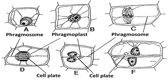
Fig: Division of highly vacuolated cells.
A, cell in non-dividing state; B, nucleus in prophase and located in the middle of the cell;
C, nucleus in early anaphase; literally mitotic spindle is connected to parietal cytoplasm by a cytoplasmic layer the phragmosome; D, daughter nuclei in telophase, the barrel shaped spindle between the nuclei is the phragmoplast; cell plate appears in its equatorial plane; E, cell plate intersects one of the walls of the mother cell F, cell division is completed and cell plate occupies the former position of a phragmosome.