Occurrence
Polypodium is a perennial herb. It is found mostly in temperate regions. It has worldwide distribution. Mostly is attached to some rocks. But some forms are epiphytic.
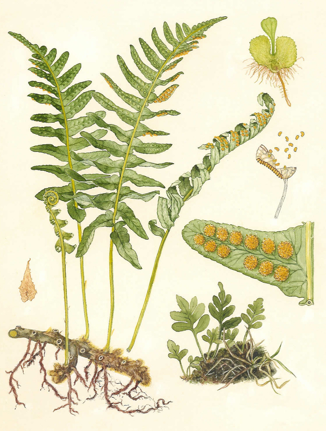
Fig: Polypodium
General structure
The plant body is sporophyte. Plant body is divided into rhizome. leaves and roots.
1. Rhizome:
It forms the main stem of the plant. Rhizome is rounded, underground. But its apex is erect. It has very few blanches. It is covered with persistant leaf bases and hairs.
2. Leaf:
The leaves are pinnately compound or simple. In compound leaf, the leaf has leaflets or pinnae. Leaves are lobed frond like. They have long stalked petiole. The leaves are arranged spirally. The form simple reticulate or dichotomous venation. The young leaves show circinate vernation.
3. Roots:
They have adventitious roots. These roots arise from the lower surface of rhizome.
Internal structure of Rhizome
In cross section rhizome is composed of epidermis, cortex and stele. Epidermis is outer most covering. It is without stomata. Cortex is wide and it is composed of parenchymatous tissues. Canal are absent in it. Stele is present in the inner side. It is covered by endodermis and pericycle. Polypodium has polystelic protostele. Each protostele has concentric vascular bundles. The xylem are exarch (protoxylem lies on the periphery of metaxylem).
Internal structure of leaf
Leaflets or lamina of leaf is covered by upper and lower epidermis. Epidermis has a layer of cutin. Lower epidermis has stomata. Mesophyll tissues are present between two epidermises. Mesophyll tissues are differentiated into palisade and spongy mesophyll. The leaf has collateral and concentric vascular bundles.
Internal structure of root
Root has simple internal structure. It has outer epidermis, cortex and stele. Stele is protostcle and diarch.
Sporangium
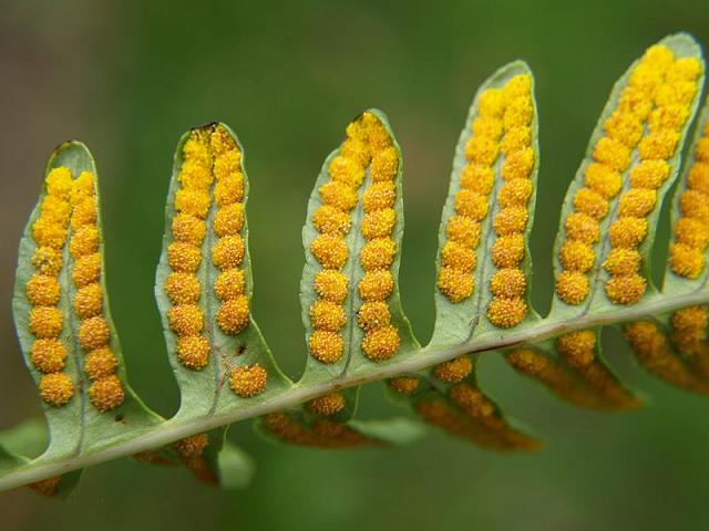
Fig: Sori on underside of leaf
Some leaves bear sporangia. They are called sporophylls. Sporophylls are foliage leaves. Sporangia are present in groups called sori. Sori are borne on the undersurface of vein of the leaves.. Sori are oval in shape. Each sorus is naked (without indusium).
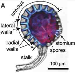
Each sorus has a group of stalk sporangia. A capsule is present on the stalk. The capsule is lenticular or biconvex. The jacket wall of capsule is single layered. This wall is differentiated into thick wall cutinized annulus and thin wall stomium. A large number of spores are present inside the capsule. Tapetum is two layered present inside the wall.
Polypodium is a homosporous. Number of spores per sporangium are 64. Spores are small, dark brown and oval. The wall of spore is composed of intine and exine. The wall ruptures at stomium during dehiscence of capsule.
Sporangium
Development of the Sporangium
I. Each sporangium develops from a single superficial cell. This cell enlarges to form an outgrowth. This cell cuts off a small cell at the base. It itself becomes the sporangial initial.
2. The sporangial initial divides transversely to form lower cell and upper cell. The lower cell is the stalk cell. The upper cell is the sporangial cell.
3. The stalk cell divides to form two celled thick stalk. The sporangial cell divides to cut off three cells at the periphery. It itself becomes tetrahedral central cell.
4. This central cell cuts off another peripheral cell at the tip. The four peripheral cells thus produced. They become jacket initials. These cells divide to produce a single layered wall. The central tetrahedral cell is the archesporial initial.
5. The archesporial initial cuts off another set of small peripheral cells. This second set of peripheral divides to produces tapetum around the archesporium. The archesporial cells undergo three or four divisions producing spore mother cells. The tapetal cells provide nourishment to the developing spores.
6. Each spore mother cell undergoes meiosis and gives rise to four spores.
Gametophyte of prothellus
The spore germinates to produce monoecious gametophyte. Gametophyte is surface living. It is differentiated dorsoventrally. It has an apical notch. It is green and heart shaped. The ventral surface of prothellus has many rhizoids. Many antheridia are present among the rhizoids toward the ventral side of mature prothellus Many archegonia are present near the apical notch.
Antheridia
The antheridia are slightly projected on the surface. They are spherical or oval in shape. Each antheridium has a layer of jacket cells. Antherozoid mother cells are present within the jacket. Sperm mother cells are changed in spirally twisted multiflagellate sperm or antherozoids.
Development of Antheridium
4. Each antheridium develops from a single superficial cell. This cell divides to form upper central cell and lower first ring cell.
5. The central cells divide to form outer jacket cell and primary androgonial cell.
6. The jacket cell divides to form a cover cell and second ring cell. The first and second ring cells and cover cells form the wall of antheridium. The primary androgonial cell undergoes several divisions to produce a mass of androcytes or antherozoid mother cells. Each androcyte changes into antherozoid.
Archegonium
The archegonia are also very numerous. Their neck is sunken in the prothellus. Each archegonium is flask shaped. It consists of curved neck and a venter. Neck is made up of several neck cells and one neck canal cells. Venter has single venter canal cell and oosphere. Development of the Archegonium
1. Each archegonium develops from a single superficial cell near the growing apex. This cell enlarges to form a small basal cell and large upper cell.
2. The basal cell divides to form venter. The upper larger cell by two transverse divisions produces a row of three cells.
3. The upper cell is the primary cover cell. It divides to form four neck initials. Neck initial divides to form neck. The middle cell is the neck canal initial. It forms neck canal cell. The lower most cell is the primary ventral cell. It divides to produce an oosphere and a ventral canal cell.
Fertilization
At the time of fertilization, the ventral canal cell and the neck canal cell disintegrate and form mucilaginous mass. This mass oozes out of the neck of archegonium. The antherozoid is attracted to the mucilage. A large number of antherozoids reach the base of the archegonium. But only one of them fuses with the oosphere to form oospore.
Development of the Embryo
1. The oospore increases in size. It divides to produce eight cells or octants. The four upper cells become epibasal cells. The four lower cells become hypobasal cells.
2. The two epibasal cells divide to form first leaf or cotyledon. The other two epibasal cells divide to form stein. The two hypobasal cells give rise to the first root. The other two hypobasal cells produce the foot. Foot penetrates into the tissue of the protballus.
3. The primary root grows for sometime under the prothallus. It then enters the soil and absorbs nutrients. Cotyledonary leaves are simple. The stem apex produce more leaves. Te stem apex grows horizontally in the soil forming the rhizome. Prothallus ultimately disappears.
Alternation of Generation
Polypodium shows a regular alternation of sporophytic and gametophytic generations. Both generations are independent. Sporophyte produces the haploid spores by meiosis. The spores germinate to form haploid prothallus or gametophyte. Prothellus is monoecious. It produces antheridia and archegonia. The union of antherozoid and oosphere produces diploid oospore. Oospore germinates to form diploid sporophyte.
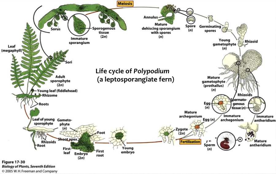
Fig: Life cycle of polypodium