A special type of cell division, which reduces the number of chromosomes to half in daughter cells as compared to parent cell. It always occur in diploid cells (2n) or tetraploid cells (4n).
In animals it takes place at the time of gamete formation (gametogenesis), while in plants at the time of spore formation (sporogenesis).
Each diploid cell after meiosis produce four haploid cells (n), because it involves two consecutive divisions after single replication of DNA.
DIVISIONS OF MEIOSIS
There are two divisions of meiosis:
- Meiosis-I
- Meiosis-II
MEIOSIS-I
It is the reduction division, which is further divided into sub-stages like Prophase-I, Metaphase-I, Anaphase-I, Telophase-I.
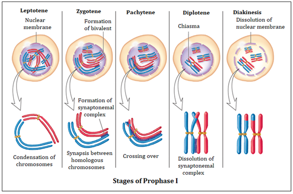
1.PROPHASE I
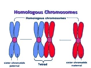
This is very prolonged phase, and differs from the prophase of mitosis, because in this chromosomes behave as a homologous pairs. Each diploid cell has two chromosomes of each type, one member from each parent, because of fusion of male and female gametes. Each chromosome has two chromatids, because chromosomes have been replicated during interphase. These similar but not necessarily identical chromosomes are called homologous chromosomes. Prophase I further consists of the following sub-stages
-
LEPTOTENE
(Gr. thin thread)
(i) The chromosomes become visible, shorten and thick due to condensation.
(ii) The size of the nucleus increases and homologous chromosomes start getting closer to each other.
-
ZYGOTENE
(Gr. paired thread)
(i) During this phase pairing of homologous chromosomes called synapsis.
(ii) This pairing is highly specific, but with no definite starting points.
(iii) Each paired but not fused, complex structure is called as bivalent or tetrad.
iii. PACHYTENE (Gr. thick band)
(i) The pairing of homologous chromosomes is completed. Chromosomes become more thick. (ii) Each bivalent has four chromatids, which wrap around each other.
(iv) Non-sister chromatids of homologous chromosomes exchange their segments due to chiasmata formation, during the process called crossing over. In this way, reshuffling of genetic material occurs, which produces recombinations.
(v) Pachytene may last for days, weeks or even years, whereas leptotene and zygotene can last only for few hours.
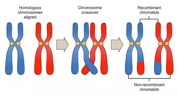
Fig: Crossing over between the non-sister chromatids of homologous chromosomes
(iv) DIPLOTENE (Gr. double band)
(i)The paired chromosomes repel each other and begin to separate.
(ii) Separations however, is not complete, because homologous chromosomes remain united by their point of interchange (chiasmata).
(iii) Each bivalent has at least one such point, the chromatids otherwise are separated.
(v)DIAKINESIS (Gr. through motion)
(i) The condensation of chromosomes reaches to its maximum.
(ii) At the same time separation of homologous chromosomes (started during diplotene) is completed, but still they are united at one point, more often at ends.
(iii) Nucleoli disappear.
-
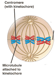
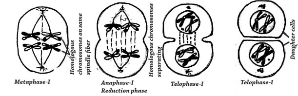
METAPHASE I
Nuclear membrane disorganize at the beginning of this phase. Spindle fibers originate and the kinetochore microtubules attach to the kinetochore of homologous chromosomes from each pole and arrange bivalents at the equator. The sister chromatids of individual chromosome in bivalent behaves as a unit.
-
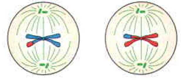
ANAPHASE I
The kinetochore microtubules contract and the spindle or pole fibers elongate, which pull the individual chromosome (each having two chromatids) towards their respective poles. It may be noted here that in contrast to anaphase of mitosis, sister chromatids are not separated. This is actually reduction phase, because each pole receives half of the total number of chromosomes.

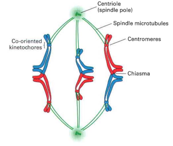
Fig: Cohesion between homologous chromosomes in meiosis I metaphase. Connections between chromosomes during meiosis I are most easily visualized in organisms with acrocentric centromeres, such as the grasshopper. The kinetochores at the centromeres of sister chromatids attach to spindle microtubules emanating from the same spindle pole, with the kinetochores of the maternal (red) and paternal (blue) chromosomes attaching to spindle microtubules from opposite spindle poles. The maternal and paternal chromosomes are attached at the chiasmata formed by recombination between them and the cohesion between sister chromatid arms that persists throughout meiosis I metaphase. Note that elimination of cohesion between sister chromatid arms is all that is required for the homologous chromosomes to separate at anaphas
-
TELOPHASE I
Nuclear membrane reorganizes around each set of chromosomes at two poles, nucleoli reappear, thus two nuclei each with half number of chromosomes are formed.
Later on, cytoplasm divides thus terminating the first meiotic division. It is also to be noted that chromosomes may not de-condensed during this state.
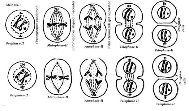
 MEIOSIS-II
It is actually equatorial division just like the mitosis. It is further divided into sub-stages like Prophase II, Metaphase II, Anaphase II, and Telophase II.
After telophase I two daughter cells experience small interphase, but in contrast to interphase of mitosis, there is no replication of chromosomes. The interphase of meiosis lack G2 phase as well. The stages of meiosis are just like the respective phase of mitosis.
PROPHASE II: The chromosomes condense and mitotic apparatus is formed.
METAPHASE II: The chromosomes arrange at equator.
ANAPHASE II: The individual/sister chromatids move apart.
TELOPHASE II: Ultimately, four nuclei at the respective poles of two daughter cells (formed after meiosis-I) are formed.
Cytokinesis takes place and four haploid cells, with half of the number of chromosomes (chromatids) are formed.